Click play icon ▶️ to listen (transcript follows below):
As a sought-after medical imaging industry thought leader, Mike Harsh sat for a recorded interview by the Radiological Society of North America that was broadcast in the US to a national radio audience.
In the recording, Mr. Harsh notes that over the last 40 years, medicine has been narrowly limited to just five ways of imaging the human body: X-ray, CT, MRI, ultrasound, and PET/SPECT. Continuing, Mr. Harsh introduces magnetic relaxometry, and speaks with great optimism and interest about the new technology’s sensitivity, specificity, and potential to improve clinical outcomes by detecting cancer much earlier.
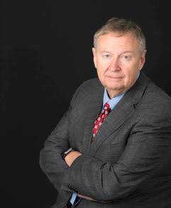
Mike Harsh is a forty-year veteran of the healthcare diagnostic imaging industry and a member of Imagion Biosystems’ Board of Directors. Mr. Harsh has served as Vice President and Chief Technology Officer for GE Healthcare and as the Global Technology Leader of Imaging Technologies at GE Global Research, where he led the organizations research on X-Ray, CT, MRI, Ultrasound, Nuclear Medicine, PET, Optical Imaging, and computer visualization/image analysis systems.
Read more about Mike Harsh at LinkedIn.
Interview Transcript:
RSNA: We’ve got x-ray and MRI and a whole bunch of things like that, but what else is new?
Mike Harsh: Well, this is what’s exciting for me. After being in this field almost 40 years now, there’s really only five ways of imaging the human body that are practical. X-ray, CT, MRI, ultrasound, and the functional modalities like PET and SPECT. I’ve had a chance now in the last several years to really work on a new one, which is magnetic reflexometry where we use magnetic nanoparticles that are injected attached to cancer cells and we can image those things with a functional imaging map, and no radiation.
RSNA: Now, how does that vary in what you’re going to find out?
Mike Harsh: We’re able to detect very specific cancer cells when there’s only tens of thousand of cells recruited. Since there’s no pan-cancer antibody, we take the iron oxide nanoparticles, we functionalize these with a specific cancer antigen that we’re after. Once it attaches, we have something that has extremely high sensitivity and specificity so we can detect cancer much earlier than is possible today.
RSNA: Okay. So once you know it’s there, then do you go to some of the more standard treatments or do you do anything different that way?
Mike Harsh: No, this’ll then fold right into standard treatments. However, when you look at clinical care pathways today, we’re sure that based on being able to detect these cancers early, we can change the clinical care pathways for much better outcomes when we can detect this cancer much earlier.
RSNA: Now, because this is relatively new, where can people find this sort of treatment?
Mike Harsh: It’s not out there yet. It’s just now starting to get into the clinical trial phase. So there’s a lot more work to do yet. But I just wanted to bring it up because there hasn’t really been anything new, when I look at ways of imaging, until recently, and this idea of using magnetic particles in some instrument, whether I detect the magnetic particles directly or through this reflexometry measurement is new. And I think it’s going to be exciting and could change medical radiology practice going forward.
