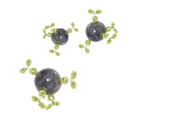
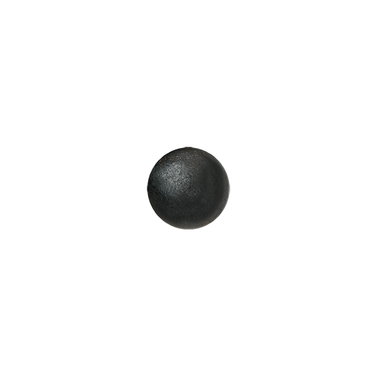
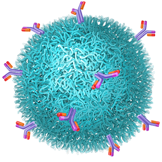
Every year cancer kills more than 9 million people globally. And each year billions of dollars are wasted and patients’ outcomes are compromised due to limitations in diagnostic imaging.
Our mission is to change the way we look at cancer, launching the next generation of molecular imaging. Today‘s gold standard provides limited information and visibility resulting in late diagnosis when tumors have grown big enough to be detectable, which is often too late.
MagSense® Imaging Agent Technology can change that.
Discreet control of uniformly sized Superparamagnetic Iron Oxide Nanoparticles (SPIONs)- Fe3O4.
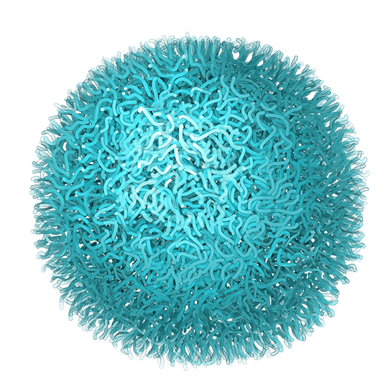
Range of biocompatible coatings to improve vary bioavailability and provide stability in physiologic conditions.
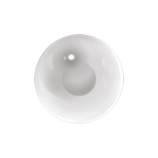
Minimizes opsonization in biological applications.
Antibodies, peptides or cancer specific small molecules provides specificity for targeting tumor cells.
MagSense® nanoparticles result in unique and differentiable image contrast when imaged by MRI.
Bio-safe magnetic nanoparticles are attracted to the tumor and detected.
Click video above to watch
Targeting molecules affixed to the nanoparticles, ensure high specificity for the cancer, and cause the nanoparticles to find and bind to tumor cells.
Most cancers are asymptomatic, growing unnoticed and undetected until the tumor has grown large enough (many millions of cells) to cause identifiable symptoms. Despite advances made in medical imaging technologies, today’s gold standard still fails to detect small and / or metastatic tumors. Additionally, current medical imaging methods cannot distinguish between benign growths and malignant cancers. This results in missed cancers, intrusive biopsies and surgeries, unnecessary treatment, and needless patient anxiety.
In this era of ever-improving precision oncology, Imagion Biosystems’ molecular MRI may provide novel insights into the presence and extent of disease, helping clinicians and patients optimize treatment plans and extend lives.
While newer blood-based testing, such as the detection of circulating tumor cells or nucleic acids, may provide earlier identification of the presence of cancer, such blood-based testing will necessitate a complementary technology to locate and stage the disease.
Enlarged, ovoid axillary lymph node
Tumor Involved?
Homogeneous Hypointensity by Normal Lymph Nodes
Heterogeneous Hypointensity in Suspicious Lymph Nodes
Enlarged axillary lymph node with replacement of the normal fatty hilum
Tumor Involved?
Uniformly darkened lymph node suggests
Benign/Normal
Heterogenous hypointensity and slightly darkened cortex confirms
Metastatic disease
Or if you’re looking to become a strategic partner to help design and drive clinical development, learn more about our collaboration opportunities.