 In our September newsletter we announced that researchers at the University of Sydney had received an industry collaboration grant to investigate the use of our nanoparticles as an MRI contrast agent. We’ve been very pleased with the progress of their research which has shown that the superparamagnetic properties of our nanoparticles provide excellent contrast for MR imaging.
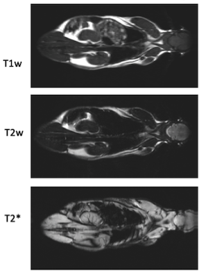
The images here (courtesy of Univ Sydney) show the presence of PrecisionMRX nanoparticles in various organs of interest. These images show how use of high-quality iron oxide nanoparticles enhances the contrast in a common type of MRI scan known as T2* weighted MRI.
With more than 50,000 clinical MRI scanners installed around the world, and known safety issues with use of gadolinium, our bio-safe nanoparticle could open the opportunity to compete in the $2 billion MR contrast agent market.
Magnetic Resonance Imaging (MRI) produces images noninvasively by using radiofrequency waves to probe the magnetization of protons (i.e. hydrogen atoms) in the constituent water molecules of various tissues within our bodies. The contrast in an MRI image is a result of variation in magnetization between hydrogen atoms in different local environments, e.g., the hydrogen bound in our fat compared to the hydrogen in our blood. In a typical MRI scan, the same image is acquired multiple times, with different system parameters yielding dramatic contrast changes between each image. Despite this, there is often insufficient contrast between tissues to distinguish them in MRI. A tumor in the breast may be invisible if the surrounding muscle and fat provides a similar MRI signal.
To overcome these challenges, scientists have developed contrast agent based imaging techniques that modify the MR signal in the area where they accrue. Our nanoparticles were chosen by researchers at the University of Sydney because when inside an MRI scanner, they behave like nanoscale bar magnets, with an incredibly concentrated magnetic field. These miniscule pockets of super-strong magnetism produce a darkening of the MR image when imaged with conventional MRI sequences. When directed to regions of interest, such as a tumor microenvironment, our nanoparticles could reveal previously unseen structure in the MR image.
Iron oxide nanoparticles were used in clinical MRI up until about 10 years ago before being gradually replaced by gadolinium-based contrast agents. However, recent concerns over long term neurotoxicity of gadolinium has reversed this trend with clinicians increasingly seeking bio-safe alternatives. Presently, there is a shortage of iron-oxide-based products approved for clinical use, presenting an opportunity for our PrecisionMRX nanoparticles once cleared by regulators such as the U.S. Food and Drug Administration (FDA).

Imagion Biosystems’ IND Application for MagSense® Phase 2 Clinical Trial Clears FDA Review
Imagion Biosystems, Ltd. (ASX: IBX) has announced that the US Food and Drug Administration (FDA) completed its review of the company’s investigational new drug (IND)