This article represents the viewpoint of management.
 When MagSense technology reaches the clinic, it will join a distinguished pantheon of time-honored cancer detection imaging methods, each with its own advantages and limitations. Among these, perhaps the most well-known is X-ray imaging.
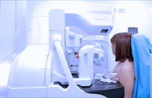
X-ray imaging works by passing ionizing radiation through the body, which is positioned between the radiation source and a detector such as X-ray film or a solid-state semiconductor panel. X-rays pass readily through air and are attenuated to varying degrees by fluids, soft tissues, and bone. Bone and other calcified structures strongly attenuate X-rays, and are visualized as areas of lighter exposure.
Although X-ray imaging cannot characterize tissues at a molecular level, it is used in cancer medicine to visualize abnormal calcifications and areas of denser-than-normal tissue, which can indicate the potential presence of a tumor. Mammograms are the most commonly recognized form of X-ray technology used in cancer medicine, but their usefulness is limited by poor results with dense breast tissue and inability to differentiate benign from malignant tumors.
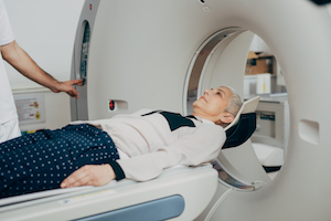 X-ray imaging, applied in a specific manner, makes CT scanning possible. In CT scanning, the patient is placed on a horizontal support inside a circular bore that passes a rotating beam of X-rays through the anatomical region of interest. Using an array of detectors within the bore, the CT scanning instrument collects imaging data from X-rays as they penetrate the patient. The resulting data are assembled by computer to create a series of cross-sectional images. This give clinicians many angles from which to view an abnormality. Contrast agents used in CT scanning, including iodinated dyes, attenuate X-ray radiation and therefore enhance visualization of structures that can temporarily contain the dyes, such as blood vessels and intestines.
All X-ray based imaging methods, including CT scanning, expose patients to low levels of ionizing radiation, a known carcinogen. Clinicians must consider this risk relative to the diagnostic value of the information to be obtained by the procedure. In CT scanning, the use of contrast agents carries an additional risk of adverse reactions, which may include rash, nausea, or more serious events.
Unlike X-rays and CT scans, MagSense imaging uses antibody recognition, the same technology that is used in pathology laboratories to analyze biopsies, to characterize benign vs. malignant tumors at the molecular level without surgical biopsy. MagSense systems require no lead shielding and do not expose patients or operators to ionizing radiation. Imagion Biosystems plans to initiate clinical studies of MagSense technology in the near future.

Imagion Biosystems’ IND Application for MagSense® Phase 2 Clinical Trial Clears FDA Review
Imagion Biosystems, Ltd. (ASX: IBX) has announced that the US Food and Drug Administration (FDA) completed its review of the company’s investigational new drug (IND)